Understanding the Connection Between Orthodontics and Cleft Jaw Surgery
Cleft lip and palate can affect the development of the upper jaw, dental arches, bite, and facial structures in ways that require long term coordination between orthodontic and surgical care. When a patient may eventually need corrective jaw surgery, orthodontic treatment cannot be planned solely around making the teeth appear straight. The position of each tooth can influence the way the jaws fit together and can affect the surgical movements that may be needed later. Presurgical orthodontic treatment is generally designed to position teeth appropriately within their individual jaw structures so the surgeon can correct the underlying skeletal discrepancy more effectively. Research emphasizes that successful treatment for patients with cleft conditions depends heavily on communication between orthodontic and surgical specialists. For families in San Antonio, understanding this relationship can make the treatment process easier to follow and can explain why an orthodontist may intentionally avoid making the bite look completely normal before surgery.
Why Future Surgery Changes the Orthodontic Strategy
In conventional orthodontic treatment, the objective may be to establish an attractive smile and a stable bite without surgical intervention. Patients with significant cleft related jaw discrepancies can require a different strategy because the skeletal relationship between the upper and lower jaws may not be correctable through tooth movement alone. The orthodontist must therefore determine whether tooth movement should compensate for the jaw discrepancy or prepare the teeth for a future surgical correction. This distinction is important because excessive dental compensation before surgery can make it more difficult for a surgeon to reposition the jaws into the desired relationship. Presurgical orthodontics commonly involves decompensating the teeth, which means positioning them more upright over their respective jaw bases rather than continuing to camouflage the skeletal discrepancy. Our approach is therefore based on understanding the eventual surgical objective before determining how individual teeth should move.
Evaluating the Jaw Before Moving Teeth
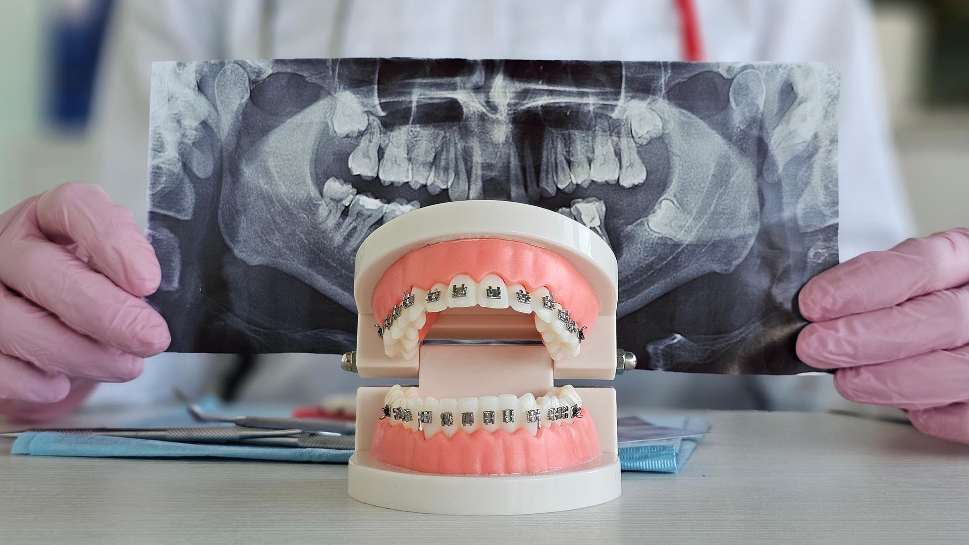
A detailed diagnosis is one of the most important parts of planning orthodontic treatment for a patient with a potential future cleft jaw surgery. The orthodontist evaluates the upper and lower dental arches, facial proportions, bite relationship, tooth inclinations, missing teeth, erupted teeth, and the condition of the bone surrounding the teeth. Imaging and digital records can help establish the three dimensional relationship between the teeth and the underlying skeletal structures. Previous cleft surgeries, alveolar bone grafting, fistulae, abnormal tooth development, and other craniofacial considerations can also influence the treatment plan. Cleft patients can have distinctive anatomical characteristics that make treatment substantially different from conventional Class III orthodontic cases. This comprehensive assessment allows the orthodontist and surgical team to establish realistic objectives before significant tooth movement begins.
Planning Tooth Position Around the Surgical Movement
One of the central principles of presurgical orthodontics is that the teeth must be positioned according to the planned movement of the jaws. A patient may initially have an underbite because the upper jaw is positioned too far behind the lower jaw, but simply moving the upper teeth forward and the lower teeth backward may camouflage the problem instead of preparing for surgery. In a surgical treatment plan, the orthodontist may instead remove dental compensation so the surgeon can advance, reposition, or rotate the jaw as necessary. This process can temporarily make the bite appear worse, even though it represents progress toward the final treatment objective. Clinical literature describes this controlled decompensation as an important part of revealing the true skeletal discrepancy and creating the appropriate conditions for surgical correction. Patients and parents should therefore understand that an interim bite does not always represent the final direction of treatment.
Managing the Upper Teeth in Cleft Cases
The upper dental arch often receives particular attention in patients with cleft conditions because maxillary growth and development can be affected by the cleft itself and by previous surgical procedures. The orthodontist may need to coordinate the width, alignment, inclination, and position of the upper teeth before the surgical phase. Teeth near the cleft area can require especially careful management because their roots and supporting bone may have unique anatomical considerations. Missing, malformed, supernumerary, or displaced teeth can further complicate decisions about spacing and tooth substitution. Presurgical planning may involve determining whether a space should ultimately be closed, maintained for a replacement tooth, or incorporated into the surgical and restorative plan. Every movement must therefore be considered in relation to the patient’s future skeletal, dental, and restorative objectives.
Coordinating the Lower Teeth With the Future Jaw Position
The lower arch also has to be positioned carefully because its relationship with the upper jaw will change when corrective jaw surgery takes place. An orthodontist may avoid making certain lower arch corrections too early if those movements could interfere with the planned surgical relationship. In moderate and severe skeletal discrepancies, research recommends concentrating early orthodontic treatment on appropriate alignment and coordination while avoiding movements that could worsen the eventual surgical relationship. The objective is not simply to create the most attractive bite at every stage of treatment but to create tooth positions that allow the jaws to be repositioned predictably. This requires controlled biomechanics and frequent reassessment as the patient develops and approaches the surgical stage. The orthodontist must also consider future growth because skeletal relationships can continue changing before the appropriate time for definitive jaw surgery.
The Importance of Timing
Timing is particularly important when orthodontic treatment is being planned around future cleft jaw surgery. Children and adolescents may require different phases of treatment because the orthodontist must account for facial growth, dental eruption, bone development, and previous procedures. Early orthodontic intervention can address specific problems without attempting to complete the entire surgical preparation years before surgery is appropriate. For patients with moderate or severe skeletal discrepancies, contemporary cleft orthodontic literature describes abbreviated early treatment that focuses on alignment, eruption management, and transverse coordination while postponing definitive presurgical preparation until closer to the appropriate surgical period. This approach can help avoid unnecessary treatment time and reduce the risk of moving teeth in ways that later need to be reversed. At each stage, the treatment plan should be reviewed against the patient’s current growth pattern and anticipated surgical requirements.
Preparing for the Surgical Appointment
As the patient approaches jaw surgery, orthodontic treatment becomes increasingly focused on creating the exact dental configuration required by the surgical plan. The orthodontist and surgeon may review digital scans, radiographs, photographs, models, and other diagnostic records to determine whether the teeth are positioned appropriately. The orthodontist may make targeted adjustments to tooth inclinations, arch coordination, midlines, and spaces before the patient proceeds to surgery. The goal is to ensure that the surgeon has an appropriate dental relationship from which to reposition the skeletal structures. Presurgical planning in cleft patients can be more complex because the treatment team must account for the three dimensional nature of maxillary deficiency as well as previous surgeries and dental differences. Careful preparation helps the surgical and orthodontic phases function as parts of one coordinated treatment plan rather than as separate procedures.
Why the Bite May Look Worse Before Surgery
A common concern for patients and parents is seeing the bite become less attractive during presurgical orthodontic treatment. This can happen because the orthodontist is removing dental compensation that previously disguised part of the underlying skeletal discrepancy. For example, teeth that were angled to make an underbite appear less severe may need to be repositioned into a more anatomically appropriate inclination before surgery. As those compensations are reduced, the overbite or underbite can temporarily become more noticeable. This change can be an intentional part of preparing the teeth and jaws for a more accurate surgical correction rather than an indication that orthodontic treatment is failing. Explaining this possibility before treatment begins can help families understand why temporary changes in appearance can be necessary for the long term result.
Working With a Craniofacial Surgical Team
Cleft jaw surgery is best approached as a multidisciplinary process because orthodontic tooth movement and surgical jaw movement directly influence one another. The orthodontist needs to understand the surgeon’s planned skeletal movements, while the surgeon needs to know whether the teeth have reached the required presurgical positions. Communication can be especially important when the patient has missing teeth, unusual eruption patterns, previous bone grafting, or other anatomical considerations. Published research consistently identifies close communication among the specialists involved in cleft care as an important factor in successful outcomes. In San Antonio, specialized craniofacial orthodontic care can involve collaboration with surgical teams so that orthodontic mechanics are considered alongside the broader facial and skeletal treatment plan. At Amini Ortho, we work with craniofacial specialists and emphasize individualized planning for patients whose orthodontic care may intersect with surgical treatment.
The Role of Digital Imaging and Treatment Planning
Modern orthodontic planning gives clinicians more information than traditional visual examination alone. Digital scans, radiographic imaging, photographs, and computerized models can help the treatment team evaluate tooth position, arch coordination, jaw relationships, and other anatomical factors. These records can also make it easier to compare changes over time and identify whether treatment is progressing toward the intended surgical objective. Digital treatment planning can be particularly useful when small differences in tooth angulation or arch coordination may influence the final surgical relationship. However, technology does not replace clinical judgment because the treatment plan must still account for growth, bone quality, previous procedures, periodontal considerations, and the patient’s individual anatomy. A carefully interpreted combination of diagnostic information allows the orthodontist to make tooth movement more deliberate and predictable.
What Happens After Jaw Surgery
Orthodontic care does not necessarily end when the jaw surgery is completed. Once the bones have healed sufficiently, the orthodontist may continue treatment to refine the bite, coordinate the arches, and address small tooth position discrepancies. Postsurgical orthodontics is generally intended to complete the occlusion rather than repeat the entire presurgical process. Research on cleft orthodontic treatment emphasizes close monitoring after surgery because healing, skeletal stability, occlusal adaptation, and functional changes can influence the finishing stage. The finishing process may include carefully selected elastic wear and small adjustments designed to establish stable contact between the teeth. Retention is also important because maintaining the achieved tooth positions becomes part of protecting the long term orthodontic and surgical result.
Why Every Tooth Movement Has a Larger Purpose
For patients with cleft conditions and potential future jaw surgery, orthodontics is not simply a process of straightening visible teeth. Each tooth has a position within a larger system involving the dental arches, jaw bones, facial proportions, soft tissues, and eventual surgical objectives. Moving a tooth too aggressively, correcting the wrong relationship too early, or failing to account for missing teeth can complicate later treatment. Conversely, carefully timed tooth movement can create a more suitable foundation for surgical correction and subsequent orthodontic finishing. Contemporary clinical guidance emphasizes that orthodontic finishing and tooth positioning should be considered alongside anticipated surgical and restorative procedures throughout cleft treatment. This is why an individualized treatment strategy is particularly important when a patient’s long term plan includes corrective jaw surgery.
Choosing Specialized Orthodontic Care in San Antonio
Families considering orthodontic treatment for a child or adult with a cleft condition should look beyond routine tooth alignment when evaluating their treatment options. A provider experienced in craniofacial orthodontics can recognize how cleft anatomy, jaw growth, previous procedures, tooth development, and potential surgery interact. Early evaluation can also help identify whether the patient is likely to require staged orthodontic care or future surgical coordination. Amini Ortho provides specialized cleft lip and palate orthodontic care in San Antonio and works with craniofacial specialists as part of complex treatment planning. The practice serves patients in West and Northeast San Antonio and offers care for children and adults with cleft and other craniofacial conditions. For families navigating a complex orthodontic journey, having a treatment team that considers future surgical needs from the beginning can provide a clearer path toward functional and aesthetic goals.
Building a Treatment Plan for the Long Term
Planning tooth movement around future cleft jaw surgery requires patience because the ideal position for a tooth today may be different from the position required immediately before or after surgery. The orthodontist must balance current dental needs with future skeletal development and the eventual surgical objective. This means that treatment may occur in carefully selected phases rather than following a single uninterrupted sequence of tooth movement. Regular diagnostic reviews allow the treatment team to determine whether the patient’s growth and dental development continue to match the original plan. The ultimate goal is to coordinate orthodontic mechanics, jaw surgery, facial development, and long term retention into one coherent treatment strategy. With careful planning and communication among the specialists involved, patients with cleft conditions can receive orthodontic care that is designed not only for today’s smile but also for the functional and structural goals of their future treatment.